e-Learning
Online Guided MRI Hip Mini Fellowship
AUD 852.50
AUD 775.00
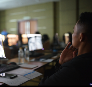
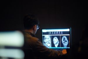
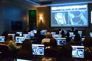
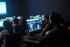
The Guided MRI HIP Online Mini Fellowship
This exciting course will cover the common abnormalities we see in reporting at work, focusing on HOW we assess a scan, WHAT to Look for, WHERE to Look and How to BEST REPORT it.
Date: This is a recurring course and runs on the first Saturday of every month
Time & Duration: 30 Days course with daily lesson released at 5:00 am SGT everyday.
Delivery method: This is an online, self-paced course
Target audience:
- Consultant and Trainee Radiologists
- Rheumatologists
- Orthopaedic Surgeons
- Sports Medicine Physicians
- and any other interested health professionals, with limited to intermediate experience in HIP MRI, who wish to improve their confidence in assessing and reporting HIP MRI
Prerequisites to attend:
- You do need to be a Health Professional and the course is not open to the general public.
- We have Consultant and Trainee Radiologists, Rheumatologists, Orthopaedic Surgeons and Sports Medicine Physicians attending our courses and the workshop will be suitable for any Medical Doctor who has an interest in or deals with MSK injuries
Course Accreditation: 30 CPD Hours by RANZCR
Free Shipping
For all orders over $400.
Got a Query?
Call us at 1300 761 006
Online Support
Contact us on online chat.
Related Products
Check items to add to the cart or
e-Learning
Online Guided MRI Foot & Toe Mini Fellowship
Ref # : V02_MR_ONL_005
AUD 852.50
AUD 775.00