e-Learning

Online Guided MRI Spine Spondyloarthopathies & Arthritis Mini Fellowship

SGD 817.50 SGD 750.00
Available
Ref.
V02_MR_ONL_007

Abstract: 

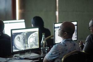
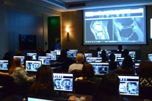
The Online Guided MRI Spine Spondyloarthropathies & Arthritis Mini Fellowship
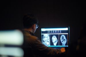
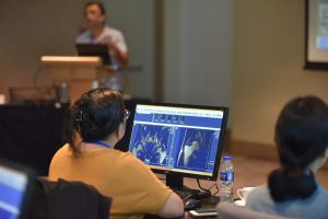
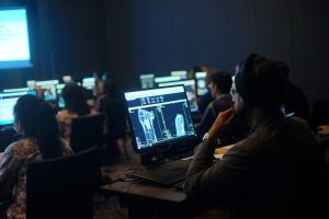
 is a structured 30-day course aimed at enhancing your diagnostic confidence, enabling you to produce clear reports that garner respect from clinicians.

This course will address common abnormalities encountered in our professional reporting work, emphasizing the methodology of scan assessment, what specific elements to search for, where to focus your attention, and the best practices for reporting. Complex information is condensed into the key knowledge required to produce a confident report.

In contrast to most online courses where you are left to navigate the material independently, our approach involves active guidance and demonstration. We will show you how to examine scans effectively, provide guidance on how to articulate your findings, and address any questions you may have to ensure all doubts are clarified. It is our priority that by the end of this Mini Fellowship, you will have the confidence to generate accurate reports.

Key Learning points: 

  • Learn to Assess, diagnose and report confidently the common and less common abnormalities you will see in MRI of the Spine Spondyloarthropathies & Arthritis at work.

  • Practical learning where you can see and assess a scan just like you do at work and apply that to the scans you report at work.

  • Guided learning with the ability to ask questions if you are unsure or need clarification at any stage of the course. You can also see all the previous questions and answers asked by other registrants.

  • Learn the relevant radiological anatomy and macroscopic pathology which are essential to make the interpretation of imaging findings easier and to report more confidently.  

  • Remembering the information is as important as learning it. There are multiple quizzes and review cases throughout the Mini Fellowship that will help you assess your understanding and retention of the learning.

  • 30 web-based learning CPD/CME Hours by RANZCR. These CPD hours are recognized by most international licensing authorities. 

Additional Information

  • We use a combination of images, videos, text and most importantly full dicoms where you can see and assess a scan just like you do at work.

  • You can ask questions if you are unsure or need clarification at any stage of the course and Dr Ravi will reply to you. You can also see all the previous questions and answers asked by other registrants.

  • There are multiple quizzes and review cases throughout the Mini Fellowship that will help you assess your understanding and retention of the learning.

  • We make sure you understand the detailed and complex radiological anatomy of the Spine Spondyloarthropathies & Arthritis essential to an accurate diagnosis. We also cover the macroscopic pathology of abnormalities in all our Mini Fellowships, which most courses dont, but it makes understanding and reporting the imaging easier.

  • There is also a pre workshop sequence of videos that will help you to learn and retain more .

  • The focus of this course, like all our courses, is for you to be MORE CONFIDENT with your diagnosis, issue Clear Reports and to have your reports respected by the referring physician.

Testimonials
Dr. Nikolas, Germany
"Systematic and logical management of the course, much better than what's covered in a book. Best radiology course I have taken so far."

Dr. Tony, India
"Systematic, excellent sample cases & case discussion, doubt clearing. Very good course never found this information in a textbook."

Topics covered:

1. SEQUENCES AND PLANNING SPINE AND SIJ IMAGING
• Which sequences do I use
• How do I plan sacrum specific scans

2. SPINE ANATOMY (BONY AND LIGAMENTOUS)
• Important anatomy I need to know

3. SIJ ANATOMY (BONY AND LIGAMENTOUS)
• Important anatomy I need to know

4. CLASSIFICATION OF SPONDYLOARTHRITIS
• The classification can be very confusing
• What do I need to know to put out a sensible report.

5. NON-RADIOGRAPHIC AXIAL SPONDYLOARTHRITIS
• If I can't see it why do I need to know about it
• Because your referrer will talk about

6. RADIOGRAPHIC AXIAL SPONDYLOARTHRITIS (ANKYLOSING SPONDYLITIS)
• SIJ Changes Acute and Chronic
• Spinal Changes Acute and Chronic
• We look at the Pathology of the changes to better understand the imaging findings

7. PSORIATIC ARTHRITIS
• Spine and SIJ Imaging features
• How do I differentiate it from other spondyloarthropathies

8. ENTHESITIS IN THE SPINE AND SIJ
• It's important to know Why it occur and What happens
• Then we look at and understand the imaging findings

9. IBD RELATED SPONDYLOARTHRITIS
• What is it and what are its features
• What to look for on Imaging

10. DISH
• Spine and SIJ Imaging features
• How do I differentiate it from other spondyloarthropathies

11. RHEUMATOID ARTHRITIS
• Cervical Spine Changes
• What do I need to know to put out a good report
- Pannus, Erosions, Instability, lnvagination, Cord compression

12. CPPD
• What is it, What to look for and How to Report it

13. BASIC CALCIUM PHOSPHATE
• What is it, What to look for and How to Report it

14. WHAT CAN MIMIC SACRO ILEITIS

15. SIJ INFECTION
• What to look for and How to Report it
• How to differentiate it from sacroiliitis

16. SIJ OSTEITIS CONDENSANS ILEII
• What to look for and How to Report it
• How to differentiate it from sacroiliitis

17. SIJ PREGNANCY RELATED ABNORMALITIES
• What to look for and How to Report it.
• How to differentiate it from sacroiliitis

18. SIJ DEGENERATION
• What to look for and How to Report it.
• How to differentiate it from sacroiliitis

About Speaker
  • Dr Ravi Padmanabhan
  • Dr Ravi Padmanabhan is the Director of Radiology Education Asia, and a Senior Consultant Radiologist from Australia, and now in Singapore. He has been teaching MSK and Spine MRI for over 10 years. His aim in the course is not just accumulating facts, but for you to be reporting more confidently at work. 

    His method of teaching is to simplify, without losing the essential things we need to know. For you to easily recognise the important anatomy, the relevant macroscopic pathology which helps to understand the imaging findings and for you to know where to look and what to look for. All of these help you to report a scan with confidence and issue reports that you are proud of and will be respected by referrers.